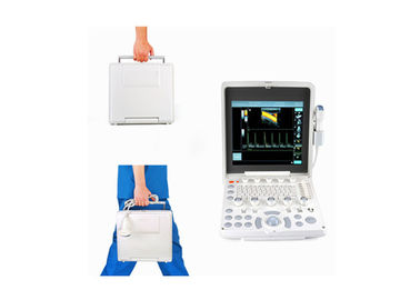
Product Details:
|
Display: | 12.1-inch LED Screen | Probe Connect: | 2 |
---|---|---|---|
USB Ports: | 2 | TCG: | 8 Segment |
B Mode: | 1024 Frames | Frame Rate: | ≥50fps |
Display Depth: | ≥30cm | High-Resolution: | 1024 *768 |
Gray Scale: | 256 | Display Language: | English |
High Light: | portable ultrasound equipment,portable ultrasound device |
Portable Color Doppler Ultrasound Scanner With 12.1 Inch Screen 2.5-10 MHz Multi-Frequency Probe
Standard Configuration
- 12.1-inch LED monitor
- Two transducer connector
- Control Panel
- 2 USB ports
- HDMI
- LAN
- Handle
- Indicators: Power/Battery/Standby
Options
-3D function
- Sony UP-897MD Printer
- One more Battery
- Gel
- printer paper
- Trolley
Ports
1.USB: 2 USB interface can be connected directly to the electronic printing device to print report and to copy data by mobile storage device
2.HDMI:Can be converted to VGA output interface,director connection to a high-resolution display and other display and other display devices,more convenient for patient to show an ultrasound image.
3.LAN: It can be used directly connected the data cable and transfer data to the internet.
Optional probe | |
5ACR60 Convex probe | R=60mm 2-5MHz |
5AL40 Linear probe | L=40mm 5-10MHz |
5AL25 Linear probe | L=25mm 5-12MHz |
5AMCR20 Micro-Convex probe | R=20mm 2-4MHz |
5AMCR11 Micro-Convex probe | R=11mm 5-8MHz |
5AMCVR Transvaginal probe | R=10mm 5-8MHz |
System Specifications | |
Display | 12.1-inch LED screen |
Probe connect | 2 |
USB ports | 2 |
Image formats | BMP, JPG, DCM, FRM,CIN |
TCG | 8 Segment |
B mode | 1024 frames |
M mode | 12.4 s |
Frame rate | ≥50fps |
Display depth | ≥30cm |
High-Resolution | 1024 *768 |
Gray scale | 256 |
Net Weight | 5.5 kg (with battery) |
Dimensions | 29.8*31.8*13.8cm |
Body markers | more than 100 Dynamic range,≤165dB |
Display Language | English |
Power | System operates via AC power |
Ports | USB: 2 USB interface can be connected directly to the electronic printing device to print report and to copy data by mobile storage device |
HDMI:Can be converted to VGA output interface,director connection to a high-resolution display and other display and other display devices,more convenient for patient to show an ultrasound image | |
LAN: It can be used directly connected the data cable and transfer data to the internet | |
Image processing | AQ sonic:Reduce random noise |
E Crystal:Adaptive speckle suppression improves contrast resolution | |
Multi-frequency probes for LD imaging modes | |
Ultra-wideband beamformer | |
Iintelligent imaging optimization technology | |
Image Zooming | |
Near and far Gain adjustment | |
With dual and duplex imaging | |
2D, Color Doppler and PW displaying on one screen in real time | |
Large capacity Li-battery | 11.1V, 8000MAH |
up to 1000 times charge-discharge,2 hours fully charged | |
Removable | |
2.5 hours regular use | |
Auto switch to battery power in case of suddenly power off | |
Display Modes | Broadband, multi-frequency imaging |
B, 2B, 4B, B+M, CFM, PDI, DPDI, PW, B+PW(Duplex) | |
B+CFM/PDI/DPDI+PW(Triplex) | |
Application | OB/GYN reporting. |
Small part analysis | |
Cardiac analysis with intimate medial thickness measurement | |
Velocity Colour to detect colour flow with PW Abdominal, Obstetrical, Gynecological, Cardiac, Small parts, Urological, Vascular, Orthopedics, MSK |
Video
Contact Person: Mrs. Helen Bo
Tel: 86-13382885910
Fax: 86-510-85572789