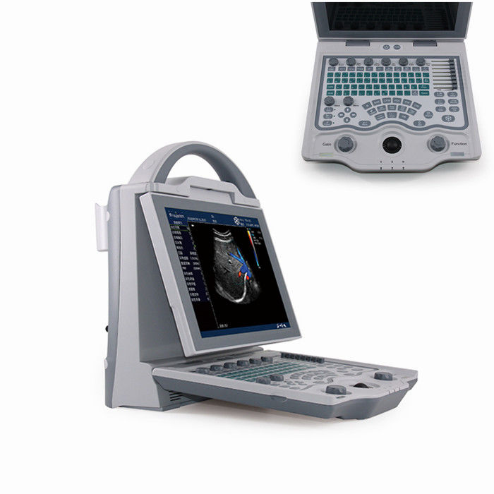
10.4 Inch Angle Adjustable Screen Portable Color Doppler Machine With 2 Probe Connector
Probe configuration
1. 3.5MHz multi-frequency abdomen convex probe
2. 6.5MH multi-frequency trans-vaginal probe
3. 7.5MHz multi-frequency linear probe
Operation Mode:B,B/B,4B,M,B/M,CFM,PDI,PW,THI
Ultrasonic imaging technology
High precise digital multi-beam formator
Dynamic frequency fusion imaging
High precise delay dynamic receiving focusing
Ultra-wideband image technique
Adaptive color artifact removal technique
Adaptive vessel imaging
Adaptive Doppler image technique
THL tissue harmonic imaging technology
Completely Independent Intellectual Property
Master Core Technology
1. Transmitted signal accuracy control
2. iMage:image optimization imaging technique
3. Adaptive color artifact removal technique
4. Linear probe independent angle deflection
5. I-station integral working station
6. Phase enhance harmonic
7. Accurate vessel image
8. Convenient and practical data management
9. Abundant measuring software
| Standard configuration |
Optional configuration |
| Main unit:1 pc |
6.5MHz multi-frequencyMicro-Convex probe:1pc; |
| probe sockets:2 |
7.5MHz multi-frequency high linear probe:1pc; |
| 3.5MHz convex probe:1pc |
Video printer |
| CD: 1pc |
Trolley |
| Reticle: 1pc |
|
Digital Specification
| Monitor |
10.4 inch color LED monitor |
| Operation Mode |
4B; M,B/M; CFM; PDI; PW; THI |
| Gray / Color scale |
256 |
| Adapter rating |
100-240V~1.2-6.0A |
| Power Frequency |
50-60Hz |
| Output of adapter |
DC12.8V 3.0A |
| Power consumption |
≤100VA |
| Main unit size |
approx. 256*150*326(mm, L*M*H) |
| Weight of main unit |
approx. 4.5kg(excluding accessories) |
| Report |
OB, Cardio, Urological report |
| U-disk capacity |
16G |
| Dual-mode TV output |
PAL/NTSC |
| Probe connector |
2 |
| Language |
English/Chinese |
| Operation mode |
B,B/B,4B mode,M,B/M mode |
| Color flow mode(CFM) |
| Power Doppler mode(PDI) |
| Pulsed-wave Doppler(PW) |
| Tissue harmonic imaging(THI) |
| Ultrasonic imaging technology |
1. High precision digitizing continued wave former |
| 2. Dynamic frequency blend imaging technology |
| 3. High precision delay point-by-point dynamic receiving focus |
| 4. Ultra-broadband imaging technology |
| 5. Automatic optimizing process image technology |
| 6. Automatic vessel imaging technology |
| 7. Automatic Doppler imaging technology |
| 8. THI structure harmonic imaging technology |
Probe Parameter
| 3.5MHz R60 multi-frequency convex probe |
| |
Scan angle |
60° |
| Frequency range |
2.0MHz-5.0MHz |
| Frequency conversion options |
2.02.53.55.0MHz |
| Max-scan depth |
≥190mm |
| Blind area |
≤5mm |
| Resolution |
Axial resolution ≤1mm, Lateral resolution ≤2mm |
| Geometrical position accuracy |
Horizontal ≤5%, Portrait ≤5% |
| 7.5MHz L40 multi-frequency linear probe |
| |
Frequency range |
5.0MHz-9.0MHz |
| Frequency conversion options |
5.06.57.59.0MHz |
| Max-scan depth |
≥60mm |
| Blind area |
≤3mm |
| Resolution |
Axial resolution ≤0.5mm, Lateral resolution ≤1mm |
| Geometrical position accuracy |
Horizontal ≤5%, Portrait ≤5% |
| 6.5MHz R11 multi-frequency trans-vaginal probe |
| |
Scan angle |
135° |
| Frequency range |
4.5MHz-9.0MHz |
| Frequency conversion options |
4.5/5.5/6.5/7.5 |
| Max-scan depth |
≥60mm |
| Blind area |
≤4mm |
| Resolution |
Axial resolution ≤1mm, Lateral resolution ≤1mm |
| Geometrical position accuracy |
Horizontal ≤10%, Portrait ≤5% |


 Your message must be between 20-3,000 characters!
 English