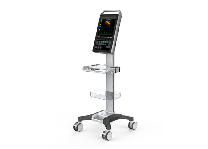
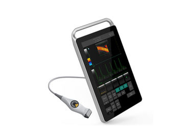
Send Message
Home ProductsColor Doppler Ultrasound Scanner

Ultrasound Scanner Equipment Color Doppler Machine With 2D Software For Human Use

Certification
China Wuxi Biomedical Technology Co., Ltd. certification
Customer Reviews
Excelente localizador de veias, excelente serviço.

—— Adilson

ผลิตภัณฑ์ที่น่าทึ่ง มีประโยชน์มากสำหรับ MSK

—— Mrs. Wantanee

Yes. The image is very good!

—— Mr. Michael Oling

Good product!

—— Dr. WAN Chun Wai Macor

Good quality and delivery in time.

—— George

Professional service

—— Fekete

I'm Online Chat Now

Ultrasound Scanner Equipment Color Doppler Machine With 2D Software For Human Use

Ultrasound Scanner Equipment Color Doppler Machine With 2D Software For Human Use
Ultrasound Scanner Equipment Color Doppler Machine With 2D Software For Human Use Ultrasound Scanner Equipment Color Doppler Machine With 2D Software For Human Use

Large Image :  Ultrasound Scanner Equipment Color Doppler Machine With 2D Software For Human Use

Product Details:
Place of Origin: China
Brand Name: BIO
Model Number: 5000M
Type: Color Doppler Ultrasound Equipments
Portable: Full digital
Payment & Shipping Terms:
Minimum Order Quantity: 1 unit
Packaging Details: 1 unit
Delivery Time: 7 working - days since getting full payment
Payment Terms: Credit card, Paypal, T/T

Ultrasound Scanner Equipment Color Doppler Machine With 2D Software For Human Use

Description
Video Output: PAL-D, S-video, NTSC, VGA, DVI Digital Scan Converter: 628× 440 × 24 Bits
Body Mark: 123 Body Marks With Probe Location Probe Frequency: 2. 0MHz~10. 0MHz
Gray Scale: 256 Color Scale: 24
Frame Rate: Max. Up To 70 F/s Scanning Area: ≥ 320 Mm
Density Of Scanning Lines: Max 256 Line/frame Cine Loop: ≥1024 Frames
Biopsy: Optional Biopsy Guide Input Power: 120VA
Continuous Working Time: >8 H
High Light:

hand held doppler

,

handheld doppler ultrasound

Ultrasound Scanner Equipment Color Doppler Machine With 2D Software For Human Use​


Function


-1 probe connector, which can be expand to 3 connectors by the probe expansion module

-18.5"high-resolution color LCD monitor

-Equipped with more than 4 beamformer function

-Equipped with THI (Tissue Harmonic Imaging), and second digital THI function

-Equipped with TSI (Tissue Specific Imaging) function

-Frequency compound imaging: adjustable under 2D and M modes

-Space compound imaging: adjustable under 2D and M modes with more than 3 levels

"-The scanning modes include 2D (2D ultrasound scanning diagnostic method), M (time motion, M mode diagnostic method), PW

(Pulsed Wave Doppler), CFM (Color Flow Mapping), PDI (Power Doppler Imaging)."

-Different frequencies are selected for 2D image and color image

-Equipped with biopsy guide line, guide by convex probe, transvaginal and linear probe

"-Equipped with bidirectional cine-loop function, gray scale cine-loop no less than 1024 frames, PW cine-loop time is no less than

100 seconds. Automatic/manual playback. The speed of the playback can be adjusted"

-Massive image storage (related with configured hard disk, no less than 500G)

-Equipped with 123 types of body marks; the probe position and scanning direction can be shown with arrow


Optional transducer


Mode Probe Frequency
CR60 Convex probe 2-5Mhz
CR11 Micro-convex probe 5-8Mhz
CR20 Micro-convex probe 2-4Mhz
L25 Linear probe 5-12Mhz
L40 Linear probe 5-10Mhz
VR10 Transvaginal probe 5-8Mhz


Specification


Monitor 18.5" LCD touch monitor
Video Output PAL-D, S-video, NTSC, VGA, DVI
Digital Scan Converter 628× 440 × 24 bits
Body Mark 123 body marks with probe location
Probe Frequency 2. 0MHz~10. 0MHz
Gray Scale 256
Color Scale 24
Frame Rate Max. up to 70 f/s
Scanning Area ≥ 320 mm
Density of Scanning Lines Max 256 line/frame
Cine loop ≥1024 frames
Biopsy Optional biopsy guide
Display Mode B, 2B, 4B, M, B+M, CFM, PDI, B+PW, B+CFM+PW, B+PDI+PW
Type of Focusing Dynamic focusing, acoustic lens focusing, launching multipoint focusing
Scanning Technique Dynamic apodization, dynamic aperture, dynamic frequency scanning, multi-acoustic beam
Pre-processing low noise preamplifier, TGC, filtering, frame average, line average
Post-processing Gamma correction, histogram, digital scan conversion (DSC), edge enhancement, noise rejection, smooth, ePure, grey scale transformation, pseudo-color, color persistence etc
Display Control Freeze/Unfreeze, Left-right reverse, Up-down reverse, Polarity reverse, picture rotation (90°/180°/270°), color reverse, inverse-frequency spectrum, pseudo-color.
Control of Transformation and Adjustment on Sound Field output acoustic power, PRF, focus position, scanning angle, image frame frequency, impulse duration, depth, sampling area dimension
General Measurement 2D/CFM: Distance, area, volume (ellipse method), angle;
M: Distance, time, slope, heart rate, simplified left ventricle function, complete left ventricle function;
PW: Distance, max ventricular gradient, average ventricular gradient, time, S/D ration, blood flow rate, acceleration of blood flow, heart rate, pulsate & drag index.
Image Memory Hard disk for massive image storage, min. 10,000 images permanent stored
Character Date, time, patient name, device name, user name etc, user-defined annotation table, arrowhead and body marks
Measurement Software Software packages of Abdomen, Urology, Cardiology, Obstetrics, Gynecology, Superficial Parts, Blood Vessels, Fetal Heart, Transcranial Doppler and Orthopedic Surgery; directly form diagnostic report based on measurement results.
Input Power 120VA
Continuous Working Time >8 h
Size(3 cartons) 65*42*18cm;140*15*9cm;73*60*39cm
Weight 40kg


Ultrasound Scanner Equipment Color Doppler Machine With 2D Software For Human Use 0Ultrasound Scanner Equipment Color Doppler Machine With 2D Software For Human Use 1Ultrasound Scanner Equipment Color Doppler Machine With 2D Software For Human Use 2


Contact Details
Wuxi Biomedical Technology Co., Ltd.

Contact Person: Mrs. Helen Bo

Tel: 86-13382885910

Fax: 86-510-85572789

Send your inquiry directly to us (0 / 3000)