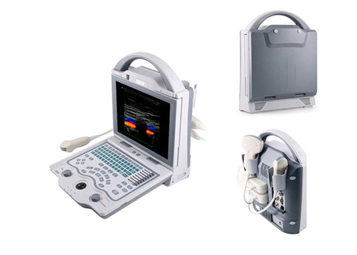
Digital Color Doppler Machine Laptop Ultrasound Scanner Diagnostic Color System
Reasonable Humanization Design
1. 10.4 inch color LED full screen display,angle adjustable
2. One-key-light keyboard function for black room use
3. One button operation including one-key storage image/ review/print,etc
4. Quick look up stored image function to improve work efficiency
5. Multifunctional knob design,achieve one-key quick adjustable in multi-mode
Powerful Data Management
1. Optimize checking image function,realize 1-2 second response
2. Intelligent user defined,one-key review and storage function
3. Neoteric internal workstation multi-plan module,quickly finish data,report,realize process multi medical histories function
Abundant Document Management
1. I-Station integral work station,achieve a key printing function With network connection;
2. Large internal 16G disk storage capacity,permanent massive storage,without loss when power off.
3. BMP format image ≥10,000framess
4. PNG format image ≥100,000 frames
Completely Independent Intellectual Property,Master Core Technology
1. Transmitted signal accuracy control
2. iMage:image optimization imaging technique
3. Adaptive color artifact removal technique
4. Linear probe independent angle deflection
5. I-station integral working station
6. Phase enhance harmonic
7. Accurate vessel image
8. Convenient and practical data management
9. Abundant measuring software
Function And Specification
| Standard configuration |
1. Main unit: 1 PC |
| 2. work station |
| 3. Reticle |
| 4. 3.5MHz multi-frequency abdomen convex probe. |
| 5. Choose between |
| 6. 6.5MHz multi-frequency trans-vaginal probe. |
| 7. 7.5MHz multi-frequency linear probe |
| Optional accessories |
1. Trolley |
| 2. Foot switch |
| 3. Thermal printer |
| 4. Ethernet switch |
| Specifications |
| Monitor |
10.4 inch color LED monitor |
| Operation Mode |
4B; M,B/M; CFM; PDI; PW; THI |
| Gray / Color scale |
256 |
| Adapter rating |
100-240V~1.2-6.0A |
| Power Frequency |
50-60Hz |
| Output of adapter |
DC12.8V 3.0A |
| Power consumption |
≤100VA |
| Main unit size |
approx. 256*150*326(mm, L*M*H) |
| Weight of main unit |
approx. 4.5kg(excluding accessories) |
| Report |
OB, Cardio, Urological report |
| U-disk capacity |
16G |
| Dual-mode TV output |
PAL/NTSC |
| Probe connector |
2 |
| Language |
English/Chinese |
| Operation mode |
B,B/B,4B mode,M,B/M mode |
| Color flow mode(CFM) |
| Power Doppler mode(PDI) |
| Pulsed-wave Doppler(PW) |
| Tissue harmonic imaging(THI) |
| Ultrasonic imaging technology |
1. High precision digitizing continued wave former |
| 2. Dynamic frequency blend imaging technology |
| 3. High precision delay point-by-point dynamic receiving focus |
| 4. Ultra-broadband imaging technology |
| 5. Automatic optimizing process image technology |
| 6. Automatic vessel imaging technology |
| 7. Automatic Doppler imaging technology |
| 8. THI structure harmonic imaging technology |


 Your message must be between 20-3,000 characters!
 English