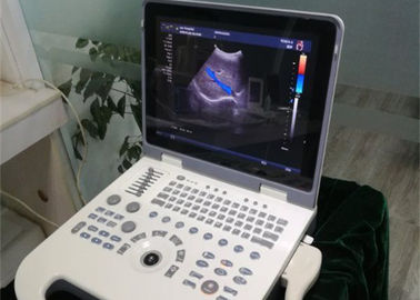
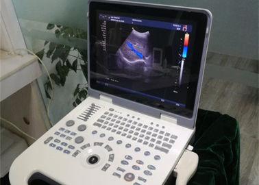
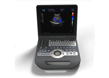
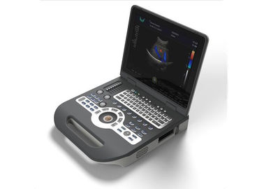
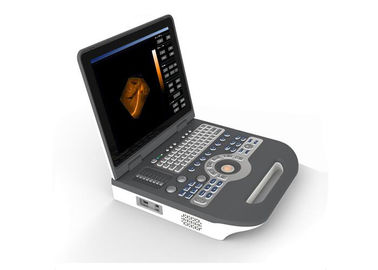
Product Details:
|
Gray Sacle : | 256 | Scanning Depth : | ≥250mm |
---|---|---|---|
Display : | 15”LCD Monitor | Mode : | B、B/B、B/M、CFM、CFM/B、PDI、PDI/B、PW |
Multi Language : | Supported | Battery: | Big Capacity Lithium |
B/D Dual-purpose: | Linear Array: B/PWD; Convex Array: B/PWD | Languages: | 5 |
Dynamic Range: | 0~120dB Adjustable | Image Storage: | ≥160G |
High Light: | hand held doppler,hand held doppler machine |
The performance requirements of Doppler spectrum mode
a). The color ultrasonic at the color Doppler spectrum mode should comply with the provisions of the;
b). Blood flow speed reading error should comply with the provisions of the table2.3;
c). Pulse wave Doppler mode sampling area cursor position should be accurate;
Displayer: 15 inch LCD color display
Running hours: ≥8h;
Input power: ≤300VA;
Host weight: about 6 kg;
Host appearance size: 370 ×382×90(length × width × height) (mm3).
Standard configuration
1 unit main unit
1 pc 3.5MHz convex probe
1 pc Power cable
Optional
1 pc 7.5MHz linear probe
1 pc 6.5MHz transvaginal probe
1 pc 5.0MHz micro-convex probe
1 pc 5.0MHz transrectal probe
1 pc thermal printer
1 pc deluxe cart
Monitor | 15 inch LCD color display |
Scanning mode | Convex,Trans-vaginal, high-frequency linear array,Micro-convex;4D array |
Modes | B,B/B,B/M,CFM,CFM/B,PDI,PDI/B,PW |
battery | Big capacity lithium |
B/D dual-purpose | Linear array: B/PWD; convex array: B/PWD |
Languages | 5 |
Gray scale | 256 levels |
Depth display | ≥250mm |
Dynamic range | 0~120dB adjustable |
One-click optimization function | Avaialble |
Self-motion optimize function | Avaialble |
Image multiplying factor | Avaialble |
Image mode | Digital beam forming, tissue harmonic imaging. |
Picture zoomfunction | Avaialble |
Image storage | ≥160G |
Image storage, video storage, cine loop | |
Frequency conversion | 2MHz-12MHz(Depending on the probe) |
Pseudo color encoding | 16 kinds can optional |
Gain adjusts | 8 segments TGC, B/M/D/C is independently adjustable |
E-zoom | magnify 2 times of real time image |
Focus position | dynamic electron focusing |
Running hours | ≥8h |
Input power | ≤300VA |
Host weight | about 6 kg |
Host appearance size | 370 ×382×90mm |
Application | Abdomen, gynecology, obstetrics, superficial organ, urologist, heart Mode and user defined model 1-4, total ten models |
Particularly Recommended | USB. VIDEO .DICOME.FOOT SW |
Output | USB Port:Our machine can connect the common printer . Used to print the measurement report page |
Patient data | Medical record management, report inquiry and printing, image video output( HDD ,USB,Optional DVD-RW),built-in ultrasound workstation |
Reporting system |
automatic report generation system, and can be full screen characters in both Chinese and English editor
|
B mode | Distance, circumference, area, volume, angle, ratio, and stenos rate |
M mode | Heart rate, time, distance, speed, ratio, etc |
Gynecology measurement | Uterus, cervix, endometrial, ovary, follicular |
Obstetrics measurement | EGA, ETD, fetal weight estimation, AFI index, OB report (including OB tables) |
Cardiology measurement | LV measurement |
Urology measurement | Prostate volume, displacement volume, bladder capacity, and residual urine output |
PW measurements | Time, speed, Heart Rate, RI, PI, etc |
Other measurement | Slice volume measurement, hip joint angle measurement |
Contact Person: Mrs. Helen Bo
Tel: 86-13382885910
Fax: 86-510-85572789