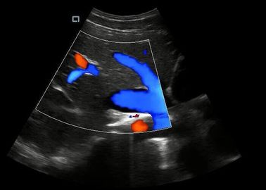
Product Details:
|
Modes: | B,M,2B,B/M,4B,C,PW, Power,DirPower,THI | Gray Level: | 256 |
---|---|---|---|
Display: | 15” LCD Monitor | Transducer Frequency: | 2-12Mhz |
Scanning Depth: | ≥300mm | Dynamic Range: | 0~120dB Adjustable |
Qopt: | One-key Optimize | Pseudo Color Encoding: | 16 Kinds Optional. |
Image Mode: | Digital Beam Forming, Tissue Harmonic Imaging | Picture Function: | Picture Zoom In And Zoom Par |
Disk Capacity: | ≥160G | E-zoom: | Magnify 2 Times Of Real Time Image |
Highlight: | hand held doppler,hand held doppler machine |
System Feature
1. 6 kinds language
2. Free hand 3D imaging
3. Multiple image modes:B/C/M/PW/PDI/Triplex
4. High-precise digital continuous beam formation
5. Dynamic Frequency Fusion Imaging Technology
6. High-precise delay point by point dynamic receiver focus
7. Ultra wideband imaging technology
8. Adaptive image optimization processing technology
9. THI tissue harmonic imaging technology
10. Precise SRI adaptive speckle noise suppression
Optional
1. Four-dimensional Imaging(4D)
2. Build battery
3. 7.5 MHz Linear array probe
4. 6.5 MHz Transvaginal probe
5. 6.5 MHz Micro convex probe
6. Lithium battery
7. 4D probe
8. Phased probe
Parameters
1. Notebook ultrasound scanner, with big capacity of lithium battery.
2. Scanning mode: Convex,Trans-vaginal, high-frequency linear array, Micro-convex;4D array;
3. Support the five languages.
4. Gray scale: 256 levels.
5. Depth display: >250mm;
6. Dynamic range: 0~120dB adjustable.
7. Image storage, video storage, cine loop, disk storage capacity >160G;
8. Image mode: digital beam forming, tissue harmonic imaging.
9. Frequency conversion: 2MHz-12MHz(Depending on the probe)
10. USB. VIDEO .DICOME.FOOT SW.
Technical Specifications
Monitor | 15' HD LED monitor |
Probe connector | 2 |
Probe holder | 2 |
USB Port | 2 |
Scanning mode | Convex,Trans-vaginal, high-frequency linear array,Micro-convex;4D array |
Modes | B,B/B,B/M,CFM,CFM/B,PDI,PDI/B,PW.CW |
B/D dual-purpose | linear array: B/PWD |
convex array: B/PWD | |
B/D/PW | real-time Triple |
Languages | 5 |
Gray scale | 256 levels |
Depth display | ≥300mm |
Dynamic range | 0~120dB adjustable |
One-click optimization function | Available |
Self-motion optimize function | Available |
Image multiplying factor | Available |
Image mode | digital beam forming, tissue harmonic imaging |
Picture function | picture zoom in and zoom par |
Disk capacity | ≥160G |
Frequency conversion | 2MHz-12MHz(Depending on the probe) |
Pseudo color encoding | 16 kinds optional. |
E-zoom | magnify 2 times of real time image |
Focus position | dynamic electron focusing |
Application Mode | Abdomen, gynecology, obstetrics, superficial organ, urologist, heart and user defined model 1-4, total ten models; |
Output Interface | USB. VIDEO .DICOME.FOOT SW. |
USB Port:Our machine can connect the common printer . Used to print the measurement report page. | |
Optional Probe | Convex,linear,Trans-vaginal,Micro-convex,4D probe,Phased probe |
Contact Person: Mrs. Helen Bo
Tel: 86-13382885910
Fax: 86-510-85572789