| Dynamic range |
0~120dB adjustable |
| Display mode |
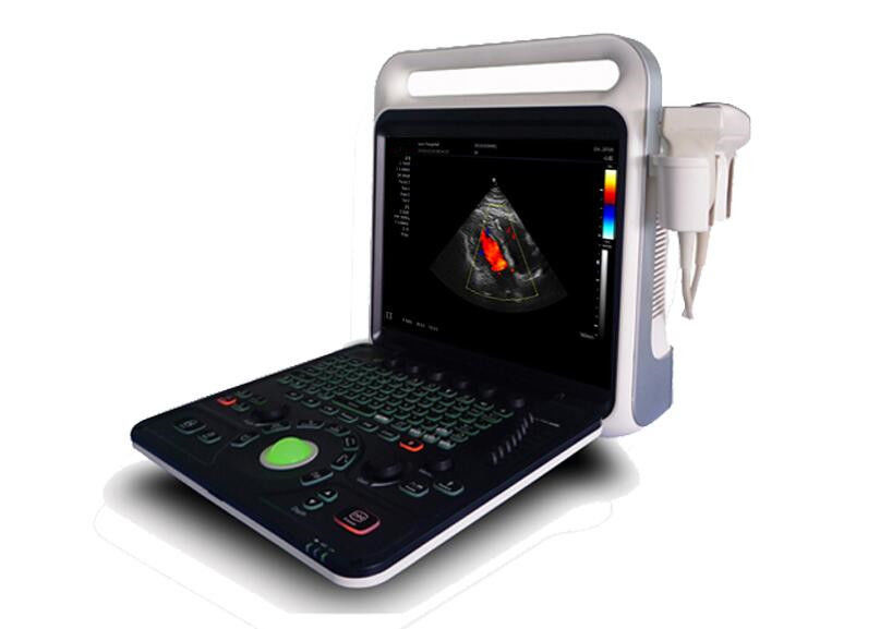
B,B/B,M,B/M,CFM,CMF/B,PDI,B/PW,CW etc mode |
| Application mode |
abdomen, kidneys, urinary system, obstetrics, gynecology, pelvic, small organ, muscle tissue, organ, breast, heart and other 11 kinds of models |
| Image mode |
digital beam forming, tissue harmonic imaging |
| IMT |
Automatic measurement and analysis vasculer intima |
| Acoustic output |
Mechanical index and thermal index real-time display |
| Acoustic power |
Step is adjustable, real-time display |
| Gray scale |
256 scales |
| Depth display |
≥250mm |
| B/D triple-purpose |
linear array: B/PW D; convex array: B/PW D |
| Pseudo color processing |
16 kinds of pseudo color encoding can optional |
| Gain adjusts |
8 segments TGC, B/M/D/C is independently adjustable; TGC curve can show and hide automatically |
| Image magnification |
picture in picture zoom in and zoom part function |
| Self-motion optimize function |
Built-in multiple check type, according to different inspection organs, preset best image check condition, reduce the adjusting operation keys |
| One-click optimization function |
preset several parameters adjusting focus on a button, a key to realize image fast optimization |
| Measurement and calculation |
--Distance, circumference, area, volume, angle, ratio, and stenos rate.
--M mode routine measurement: Heart rate, time, distance, speed, ratio, etc.
--Gynecology measurement: Uterus, cervix, endometrial, ovary, follicular
|
| Obstetrics measurement |
EGA, ETD, fetal weight estimation, AFI index, OB report (including OB tables).Cardiology measurement: LV measurement. |
| Urology measurement |
Prostate volume, displacement volume, bladder capacity, and residual urine output. |
| PW measurements |
Time, speed, Heart Rate, RI, PI, etc |
| Other measurement |
Slice volume measurement, hip joint angle measurement. |
| Image storage |
Image storage, video storage, cine loop, disk storage capacity≥160G |
| Patient data |
Medical record management, report inquiry and printing, image video output( HDD ,USB,Optional DVD-RW),built-in ultrasound workstation |
| Reporting system |
automatic report generation system, and can be full screen characters in both Chinese and English editor |
| Output interface |
SR323,USB,DICOM interface |

 Your message must be between 20-3,000 characters!
 English